Introduction
Anatomy is a branch of medical science that deals with the understanding of the structural organization of the human body. Anatomy is divided into gross/morphological anatomy, microscopic anatomy/histology, developmental anatomy/embryology and genetics. Approaches to studying gross anatomy are regional, systemic, clinical /applied, cross sectional & radiological anatomy, and includes embalming techniques of cadaver, histological and preservation techniques of human body parts are also dealt with this subject.
The Department of Anatomy was started in 1999 for the BDS course, 2006 for the MBBS course and the postgraduate MD Anatomy course was started in 2012 with 2 students. Presently the department imparts teaching and training to BDS, BSC Nursing, MSC Nursing and DMLT courses.
The department motivates young undergraduate and postgraduates to take up small research projects.
The broad goal of the teaching of undergraduate students in Anatomy aims at providing comprehensive knowledge of the gross and microscopic structure and development of human body, to provide a basis for understanding the clinical correlation of organs or structures involved and the anatomical basis for the disease presentations.
Teaching clinical anatomy with the following objectives
- Comprehend the normal disposition, clinically relevant interrelationships, functional and cross sectional anatomy of the various structures in the body.
- Identify the microscopic structure and correlative elementary ultra structure of various organs and tissues and correlate the structure with the functions as a prerequisite for understanding the altered state in various disease processes.
- Comprehend the basic structure and connections of the central nervous system to analyze the integrative and regulative function of the organs and systems. Students shall be able to locate the site of gross lesions according to the deficits encountered.
- Demonstrate knowledge of basic principles and sequential development of the organs and systems; recognize the critical states of development and the effects of common teratogens. Students shall be able to explain the developmental basis of the major variations and abnormalities
Facilities
-
Lecture Hall
Fully air-conditioned and equipped with audiovisual system
-
Demonstration Room
The 2 demonstration rooms of 75 seating capacity, equipped with LCD and white board.
-
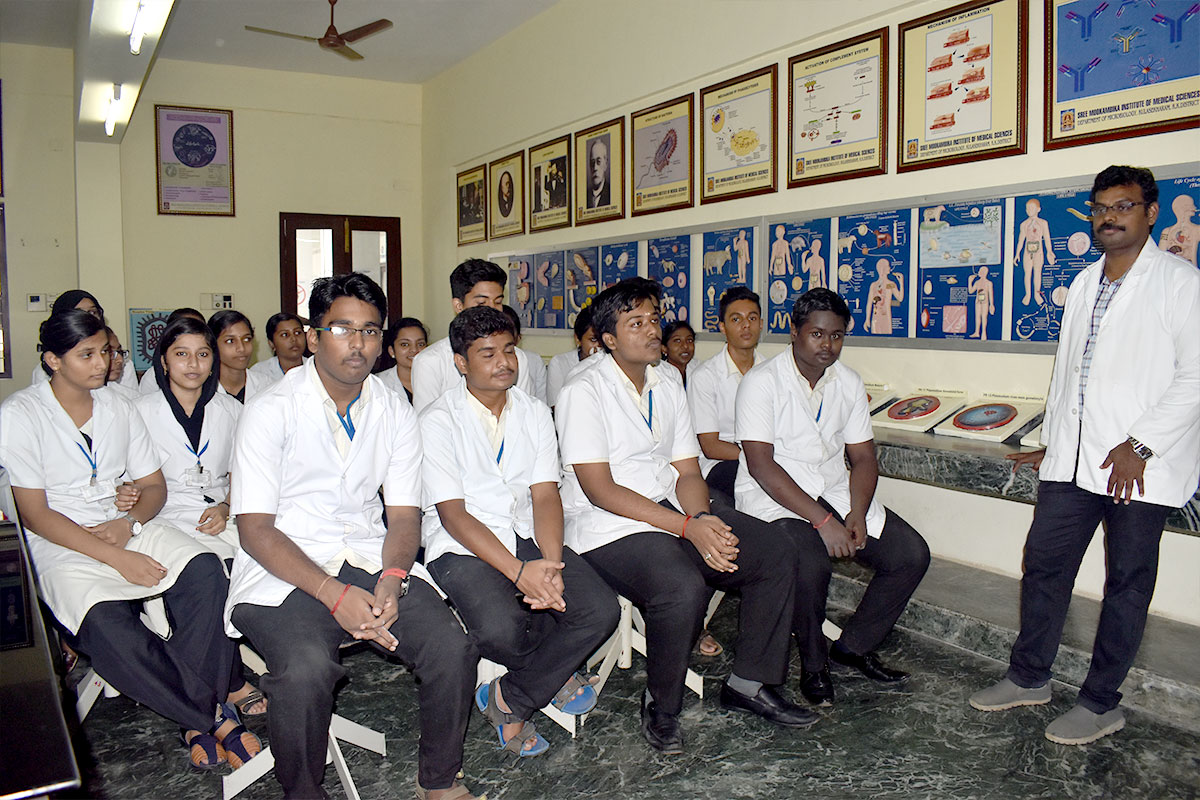
Histology Laboratory
The practical laboratory for histology accommodates 100 students and each student is provided with an individual microscope and a set of slides. The unique feature of the lab is the presence of slide projector and numerous histology charts that enables an excellent teaching learning out-come. Histology laboratory is provided with TV and video camera and LCD for projection of histology slides for small group teaching.
-
Dissection Hall
The students are motivated to develop their skills in dissection so as to understand the intricacy of human body architecture .The students are also provided prosected parts and the entire faculty interacts with students during practical session. The students are evaluated periodically by conducting notified test and terminal examination. In the dissection hall bones are displayed in a unique way for easy access to the students during dissection schedule. Also equipped with teaching aids like charts, skeleton and audiovisual aids
-
Embalming Room
Equipped with embalming machine, cold storage, 4 storage tank, bone and meat cutting machine
-
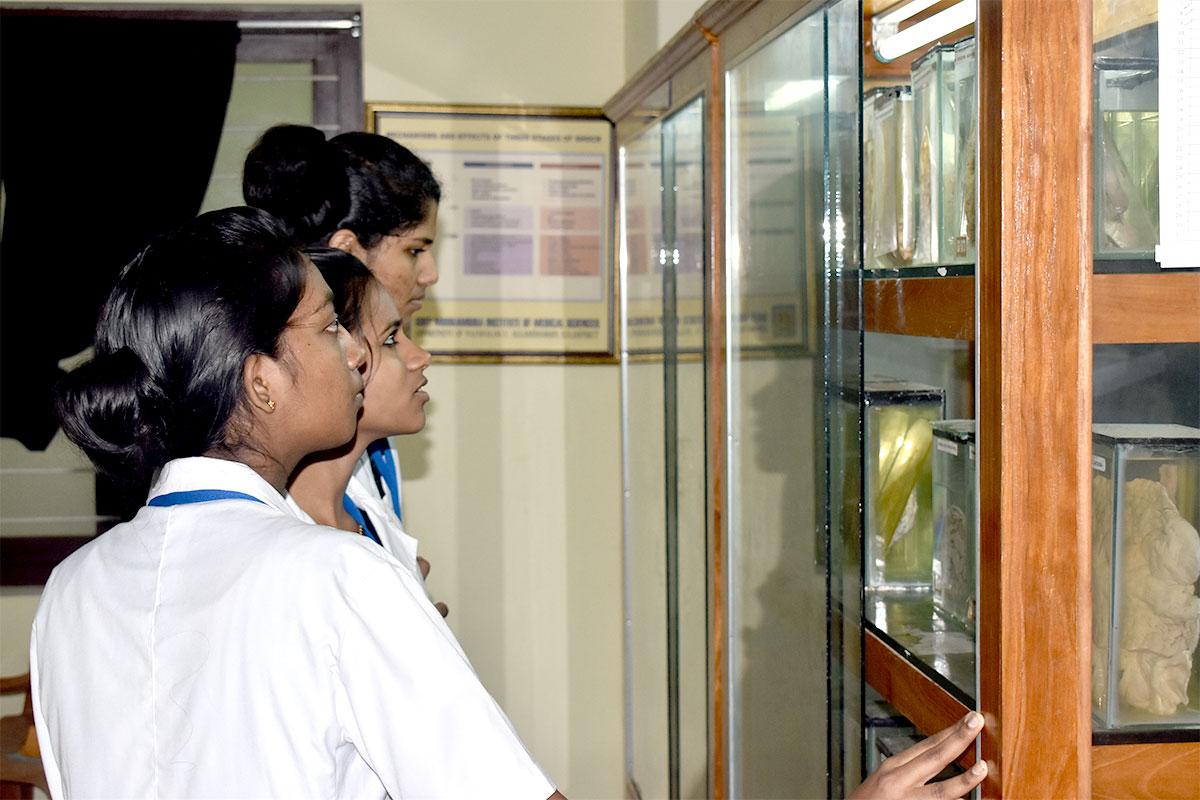
Museum
The well organized comprehensive Anatomy museum is a unique feature of the department. About 500 wet specimens are displayed region wise. Numerous charts, embryology models, skiagrams, MRI, CT scan are adequately displayed in separate sections. Transectional models, 3D models and specimens of rare congenital foetal anomalies are the added attractions to the museum.
-
Research Laboratory
The research laboratory is well equipped with binocular research microscopes, stereoscopic dissecting microscope, trinocular microscope with digital camera and egg incubator. The research laboratory provides an ideal environment for the faculty and students to carry out research activities.
-
Departmental Library
Departmental library has 176 books on Anatomy and allied subjects as reference books for the need of students/postgraduates and faculty. The library has also collections of CD’S on Anatomy for self study, improvement and assessment.
Faculty
| Professor & HOD | Dr. K. Girija Kumari |
| Professor | Dr. N. Mugunthan |
| Assistant Professor | Dr. Rathija Sreekumar Dr. Susie David |
| Tutor | Dr. Preethi Dr. Neil James Dr. Velusamy |
Horizontal Integration
The preclinical departments together plan the horizontal integration, to stress the importance of clinical and applied Anatomy
- Display study questions on the notice board weekly, pertaining to the regions covered
- At the end of a region students are given cases of the region for study, presentation analysis and discussion.
- Invite clinicians to give guest lectures and demonstrations to highlight the anatomical basis of the clinical conditions, region wise to effect clinical anatomical correlative study
Min. Working hours
Lecture hours – 100
Practical hours – 175
Total hours – 275
Evaluation
|
Theory -100 |
Practicals -100 |
|
University written exam -70 |
University written exam -90 |
|
Viva voce-20 |
Internal assessment (written) -10 |
|
Internal assessment (written) -10 |
|
Other Academic Activities
- Seminar presentation by students on relevant topics
- Group discussion as a method of problem solving Learning
- Guidance to slow learners
*Extra care and guidance is given to slow learners/ low achievers by conducting extra classes to improve their performance. We could achieve 80% results in the University Examination amongst the low achievers.
*Faculty members are engaged in small research projects and participate in national and regional conferences.